Cell: The Fundamental Unit of Life
Introduction
Every living organism—from single-celled bacteria to complex multicellular organisms like plants and human beings—is made up of microscopic units called cells. Just as bricks are the basic structural units of a building, cells are the fundamental building blocks of all living organisms.
What is a Cell? (Meaning and Definition)
A Cell is defined as the basic, smallest, structural, functional, and biological unit of life capable of independent existence and performing all essential life processes.
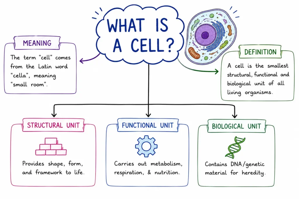
Key Elements of the Definition:
- Etymology: The word Cell originates from the Latin word cellula, which translates to “a small room” or “compartment.”
- Why is it called the Structural Unit? All living structures are constructed from cells. Cells group together to form tissues, tissues combine to form organs, organs form organ systems, and organ systems build a complete living organism:
Cells → Tissues → Organs → Organ Systems → Organism - Why is it called the Functional Unit? All fundamental life processes—such as energy production (respiration), nutrient processing (metabolism), waste elimination (excretion), and reproduction—take place inside individual cells.
- Cytology vs. Cell Biology:
- Cytology: The branch of biological science dedicated specifically to the structural study of cells under microscopes.
- Cell Biology: A broader field studying cell structure, molecular function, chemical composition, environmental interaction, and life cycles.
Discovery of the Cell and Cell Theory
The study of cell biology developed alongside improvements in optical lenses and microscopy during the 17th and 19th centuries.
1. Key Historical Discoveries
- Robert Hooke (1665): Discovered cells while examining a thin slice of bottle cork under a primitive microscope. He observed tiny, hollow compartments resembling honeycomb rooms and coined the term “Cell” (derived from the Latin word cellula, meaning “a small room”). Hooke observed dead cell walls.
- Anton van Leeuwenhoek (1674): Using an improved microscope, he became the first person to observe and describe free-living cells in pond water, including bacteria, protozoa, and human red blood cells.
- Robert Brown (1831): Discovered and named the Nucleus within orchid root cells.
- J.E. Purkinje (1839): Coined the term Protoplasm for the fluid substance present inside the cell.
- Matthias Schleiden (1838) & Theodor Schwann (1839): Formulated the classical Cell Theory. Schleiden (a German botanist) stated that all plants are composed of cells, while Schwann (a German zoologist) concluded that all animals are made of cells.
- Rudolf Virchow (1855): Expanded the cell theory by adding the famous phrase “Omnis cellula-e-cellula” (All cells arise from pre-existing cells).
2. Postulates of Modern Cell Theory
- All living organisms are composed of one or more cells and cell products.
- The cell is the fundamental structural and functional unit of life.
- All cells arise from pre-existing cells through cell division.
- The flow of energy (metabolism) occurs within cells.
- Cells contain hereditary information (DNA) passed from parent cell to daughter cell during division.
Structural Classification: Prokaryotic vs. Eukaryotic Cells
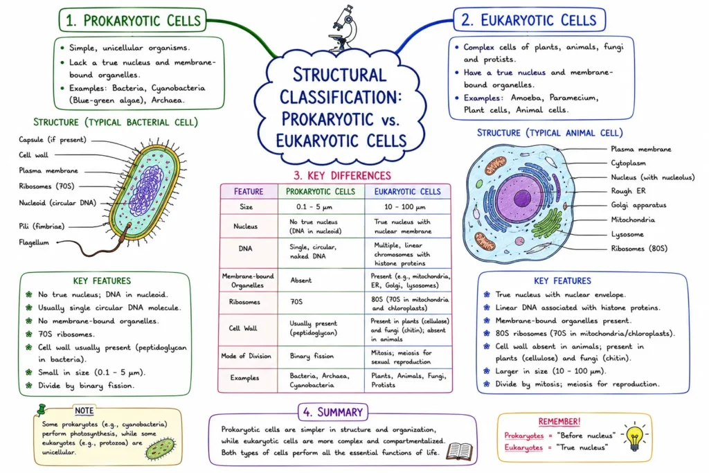
Based on internal nuclear organization and the presence or absence of membrane-bound organelles, cells are classified into two fundamental groups:
1. Prokaryotic Cells
Prokaryotic cells (from Greek pro = before, karyon = nucleus) are primitive, small, and structurally simple cells. They lack a well-defined membrane-bound nucleus and membrane-bound organelles.
- Nuclear Region: Lacks a nuclear membrane. Their genetic material consists of a single, circular strand of double-stranded DNA located directly in the cytoplasm within an undefined region called the Nucleoid.
- Organelles: Lack membrane-bound organelles (like mitochondria, endoplasmic reticulum, or Golgi apparatus). However, they contain non-membrane-bound 70S ribosomes.
- Cell Boundary: Possess a rigid cell wall made of peptidoglycan (in bacteria) surrounding the plasma membrane.
- Examples: Bacteria, Cyanobacteria (Blue-green algae), Mycoplasma (PPLO), and Archaea.
2. Eukaryotic Cells
Eukaryotic cells (from Greek eu = true, karyon = nucleus) are advanced, larger, and structurally complex cells.
- Nuclear Region: Possess a well-defined true nucleus enclosed by a double-layered nuclear envelope containing linear DNA organized into chromosomes.
- Organelles: Contain specialized membrane-bound organelles performing distinct metabolic functions.
- Ribosomes: Contain larger 80S ribosomes in the cytoplasm (and 70S ribosomes inside mitochondria and chloroplasts.
- Examples: Protists, Fungi, Plants, and Animals.
Detailed Structure of a Eukaryotic Cell
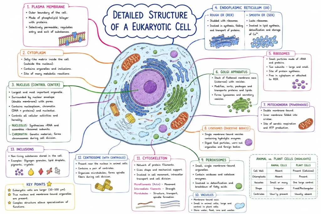
A typical eukaryotic cell consists of three main structural divisions: the Cell Envelope/Boundary, Cytoplasm containing Organelles, and the Nucleus.
1. Cell Envelope and Boundaries
A. Plasma Membrane (Cell Membrane)
The outer selectively permeable biological membrane that encloses the cellular contents, separating the interior of the cell from its external environment.
- Fluid Mosaic Model: Proposed by S.J. Singer and G.L. Nicolson (1972). According to this model, the plasma membrane consists of a quasi-fluid phospholipid bilayer with proteins embedded or floating within it like “icebergs in a sea of lipids.”
- Selective Permeability: It regulates the entry and exit of substances. Small non-polar molecules move across by simple passive diffusion, water moves by osmosis, while ions and polar molecules require active transport (expending ATP energy).
B. Cell Wall
A non-living, rigid outer layer present outside the plasma membrane in plant cells, fungi, bacteria, and algae. Animal cells completely lack a cell wall.
- Composition: Cellulose, hemicellulose, and pectin in plants; Chitin in fungi; Peptidoglycan in bacteria.
- Function: Provides mechanical support, structural shape, and protection against mechanical stress and osmotic bursting.
2. The Nucleus: Control Center of the Cell
The nucleus is the largest and most prominent organelle in a eukaryotic cell, acting as the administrative headquarters or “brain of the cell.”
- Nuclear Envelope: A double-membrane barrier perforated by tiny pores (nuclear pores) that control the passage of RNA and proteins between the nucleus and cytoplasm.
- Nucleoplasm: The semi-fluid matrix contained inside the nuclear envelope.
- Nucleolus: A dense, non-membrane-bound spherical structure inside the nucleus responsible for the synthesis of Ribosomal RNA (rRNA) and assembly of ribosomes. It is called the “ribosome factory.”
- Chromatin: An uncoiled network of thread-like nucleoprotein fibers composed of DNA and basic proteins called Histones. During cell division, chromatin condenses to form distinct rod-shaped structures called Chromosomes.
3. Cytoplasm and Cell Organelles
The Cytoplasm is the semi-fluid jelly-like matrix filling the interior of the cell between the plasma membrane and the nuclear envelope. It consists of the fluid portion (cytosol) and metabolic machinery called Cell Organelles.
A. Mitochondria: “Powerhouse of the Cell”
- Structure: Double-membrane-bound organelle. The outer membrane is smooth, while the inner membrane forms numerous deep infoldings called Cristae, which increase the surface area for chemical reactions.
- Function: Site of aerobic cellular respiration. They oxidize food items to generate energy stored in the form of Adenosine Triphosphate (ATP), earned as the “Energy Currency of the Cell.”
- Autonomy: Mitochondria are semi-autonomous organelles because they contain their own single circular DNA, RNA, and 70S ribosomes, allowing them to synthesize some of their own proteins and self-replicate.
B. Endoplasmic Reticulum (ER)
A network of interconnected membrane-bound tubules and flattened sacs spreading through the cytoplasm, connected to both the nuclear envelope and plasma membrane.
- Rough Endoplasmic Reticulum (RER): Bears ribosomes on its outer surface. It is actively involved in protein synthesis, processing, and secretion.
- Smooth Endoplasmic Reticulum (SER): Lacks ribosomes on its surface. It is the primary site for lipid and steroid hormone synthesis, as well as carbohydrate metabolism and detoxification of drugs/poisons in liver cells.
C. Golgi Apparatus (Golgi Complex)
- Discovery: Discovered by Camillo Golgi (1898) using silver nitrate staining.
- Structure: Consists of stacks of flat, membrane-bound sacs called cisternae.
- Function: The “packaging and dispatch unit” of the cell. It receives proteins and lipids synthesized by the ER, modifies them (e.g., glycosylation to form glycoproteins), packages them into vesicles, and targets them to internal or external destinations. It also forms acrosomes in human sperm.
D. Lysosomes: “Suicidal Bags of the Cell”
- Structure: Single-membrane-bound vesicular structures formed by the Golgi apparatus.
- Hydrolytic Enzymes: Filled with digestive enzymes (lipases, proteases, carbohydrates) that operate optimally at an acidic pH.
- Function: They digest foreign materials, worn-out cell organelles, and cellular debris. When a cell becomes damaged or aged, lysosomes may burst, releasing enzymes that digest their own cell—hence termed “Suicidal Bags.”
E. Ribosomes: “Protein Factories of the Cell”
- Discovery: Discovered by George Palade (1953) under an electron microscope.
- Structure: Non-membrane-bound particles composed of ribosomal RNA (rRNA) and proteins. They are found free in the cytosol or attached to the RER.
- Function: The universal site for protein translation/synthesis in all living organisms.
F. Plastids (Found in Plant Cells)
Double-membrane-bound organelles present exclusively in plant cells and algae. They are classified into three types based on pigments:
- Chloroplasts: Contain green pigments (Chlorophyll) and carotenoids. They capture solar energy to perform Photosynthesis and are referred to as the “Kitchen of the Cell.” Like mitochondria, chloroplasts contain their own circular DNA and 70S ribosomes.
- Chromoplasts: Contain fat-soluble carotenoid pigments (carotene, xanthophylls) giving red, yellow, or orange colors to flowers and fruits.
- Leucoplasts: Colorless plastids used for food storage:
- Amyloplasts: Store carbohydrates (starch, e.g., potato).
- Elaioplasts: Store oils and fats (e.g., seeds).
- Aleuroplasts: Store proteins.
G. Vacuoles
- Structure: A membrane-bound space found in the cytoplasm, enclosed by a single specialized membrane called the Tonoplast.
- Function: In plant cells, vacuoles are extremely large (occupying 50–90% of cell volume) and store water, sap, excretory products, and ions, maintaining cell turgidity and rigidity.
H. Centrosome and Centrioles
- Non-membrane-bound organelle found predominantly in animal cells.
- Contains two cylindrical structures called Centrioles arranged perpendicular to each other.
- Function: Organizes spindle fibers that separate chromosomes during cell division.
Comparison: Plant Cell vs. Animal Cell
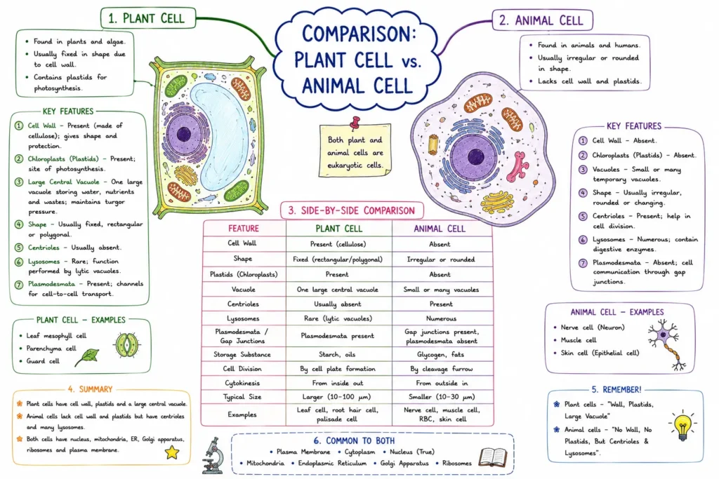
| Feature | Plant Cell | Animal Cell |
|---|---|---|
| Cell Wall | Present (composed of Cellulose) | Absent |
| Plasma Membrane | Present (inside cell wall) | Present (outermost layer) |
| Plastids / Chloroplasts | Present | Absent |
| Vacuoles | Single, permanent, central, and very large | Small, temporary, or completely absent |
| Centrosome / Centrioles | Absent in higher plants | Present (aids cell division) |
| Cilia and Flagella | Rare / Absent | Frequently present |
| Stored Carbohydrate | Starch | Glycogen |
Overview of Cell Division
Cell division is the biological process by which a parent cell divides into two or more daughter cells. It is essential for growth, repair, tissue regeneration, and reproduction.
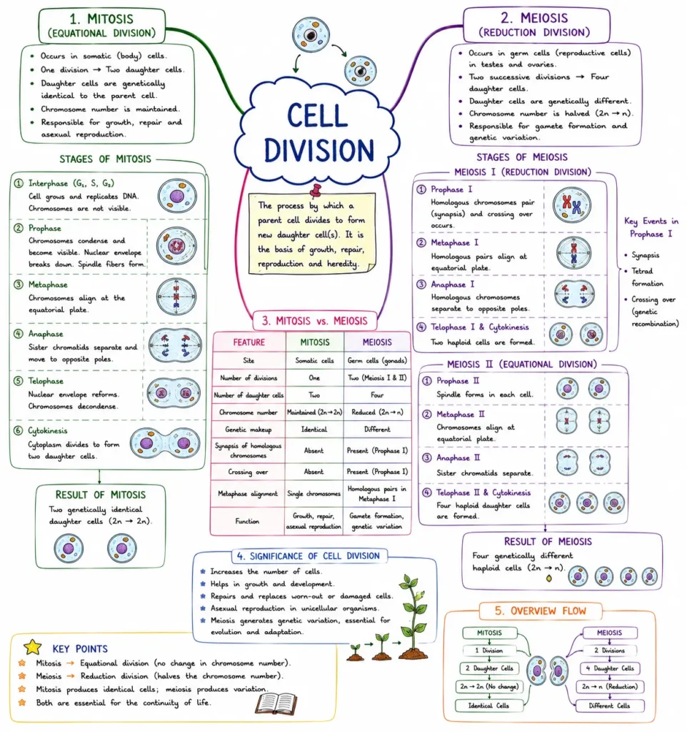
1. Mitosis (Equational Division)
- Occurs in non-reproductive body cells (Somatic Cells).
- A single parent cell (2n) divides once to form two identical daughter cells, each having the exact same chromosome number (2n) as the parent cell.
- Function: Responsible for bodily growth, tissue repair, and asexual reproduction.
2. Meiosis (Reductional Division)
- Occurs in specialized reproductive cells (Germ Cells) to produce gametes (sperm and egg).
- Involves two sequential nuclear divisions (Meiosis I and Meiosis II), resulting in four non-identical daughter cells.
- The chromosome number is halved from diploid (2n) to haploid (n).
- Function: Ensures genetic variation through crossing over and maintains constant species chromosome numbers across generations.
Quick Examination Facts
| Concept / Organelle | Key Textbook Fact |
|---|---|
| Definition of Cell | Smallest basic structural and functional unit of life |
| Etymology of ‘Cell’ | Derived from Latin cellula meaning “a small room” |
| Term ‘Cell’ Coined By | Robert Hooke (1665, observed cork cells) |
| First Live Cell Observed By | Anton van Leeuwenhoek (1674) |
| Discovery of Nucleus | Robert Brown (1831) |
| Cell Theory Formulated By | Matthias Schleiden (1838) & Theodor Schwann (1839) |
| Phrase “Omnis cellula-e-cellula” | Rudolf Virchow (1855, means all cells arise from pre-existing cells) |
| Fluid Mosaic Model Authors | S.J. Singer and G.L. Nicolson (1972) |
| Brain / Control Center of Cell | Nucleus |
| Powerhouse of the Cell | Mitochondria (Generates energy as ATP) |
| Suicidal Bags of the Cell | Lysosomes (Contain acidic hydrolytic enzymes) |
| Protein Factories of the Cell | Ribosomes (Non-membrane-bound) |
| Kitchen of the Cell | Chloroplasts (Site of photosynthesis in plants) |
| Packaging & Dispatch Unit | Golgi Apparatus (Discovered by Camillo Golgi) |
| Vacuole Membrane Name | Tonoplast |
| Semi-Autonomous Organelles | Mitochondria and Chloroplasts (Possess own DNA & 70S ribosomes) |
| Plant Cell Wall Composition | Cellulose |
| Fungal Cell Wall Composition | Chitin |
| Equational Cell Division | Mitosis (Forms 2 identical diploid cells) |
| Reductional Cell Division | Meiosis (Forms 4 haploid gamete cells) |
Conclusion
The cell is the structural, functional, and metabolic foundation of all biological existence. From primitive prokaryotic bacteria to highly specialized eukaryotic plant and animal cells, internal cellular organization dictates life processes. Understanding the structural division of organelles—including energy generation in mitochondria, protein assembly in ribosomes, structural governance by the nucleus, and cellular reproduction through division—provides the essential foundation required to study tissue structures, human physiology, genetics, and plant biology.
Cell: The Fundamental Unit of Life
Practice Quiz
| Some Important Links | |
| Download “Cell: The Fundamental Unit of Life” PDF | Click Here |